Research and Development Projects Adopted in FY2015
Spatiotemporal visualization of biopharmaceuticals by whole-body/-organ imaging with single cell resolution
Project Leader:Ueda Hiroki
Professor, Graduate School of Medicine, The University of Tokyo
Biopharmaceutical industry wishes state-of-the-art technologies such as a comprehensive and high-throughput analytical platform of in vivo dynamics of biopharmaceuticals with cellular resolution.
We have recently developed 1) whole-body/-organ clearing protocol, 2) whole-organ/-body imaging with single cell resolution, and 3) computational analysis pipeline, termed CUBIC (clear, unobstructed brain imaging cocktails and computational analysis).
CUBIC preserves the fluorescent signals from endogenous reporter proteins, and is compatible with a whole-body/-organ cell-nuclear counterstaining and a whole-organ immunostaining.
Since biopharmaceuticals can be chemically crosslinked with endogenous adjacent proteins via a conventional PFA fixation, CUBIC is potentially applicable to visualize spatiotemporal cellular distributions of biopharmaceuticals.
In this project, we aim at developing a novel analytical pipeline of in vivo dynamics of biopharmaceuticals based on the CUBIC technology, such as 1) multicolor detection of various kinds of fluorescent-labeled biopharmaceuticals, 2) superposition of biopharmaceutical distribution and nuclear/fluorescent protein/immunostained images, and 3) temporal in vivo dynamics of biopharmaceuticals.
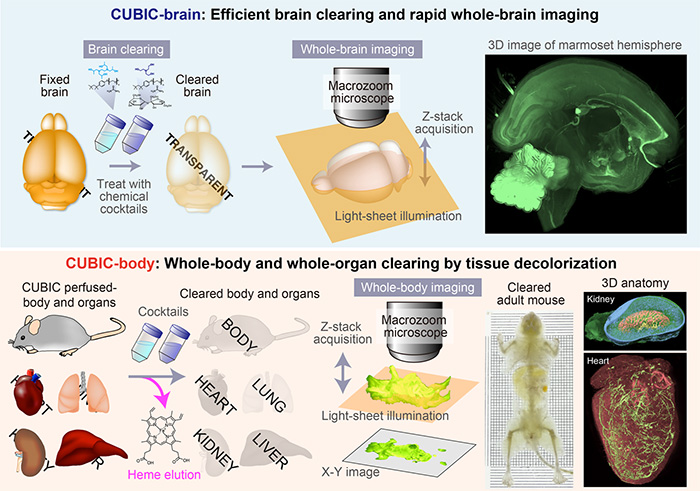
<Figure1>
Whole-brain/body clearing and imaging method, termed CUBIC (clear, unobstructed brain/body imaging cocktails and computational analysis).
Hydrophilic CUBIC cocktails significantly enhanced whole-brain clearing via effective delipidation and homogenizing mismatched RIs without signal loss from fluorescent proteins.
Additionally, an aminoalcohol in CUBIC reagent displayed highly efficient decolorization of the PFA-fixed blood by the elution of heme chromophore from hemoglobin.
Our CUBIC protocol enables rapid whole-body and whole-organ imaging with single-cell resolution by using light-sheet fluorescent microscopy.
<Figure2>
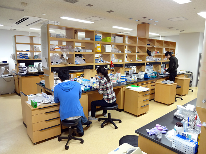
Molecular biology laboratory.
<Figure3>
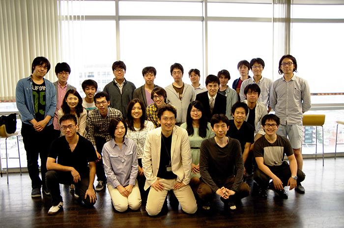
Members of the Department of Systems Pharmacology, Graduate School of Medicine, the University of Tokyo.